
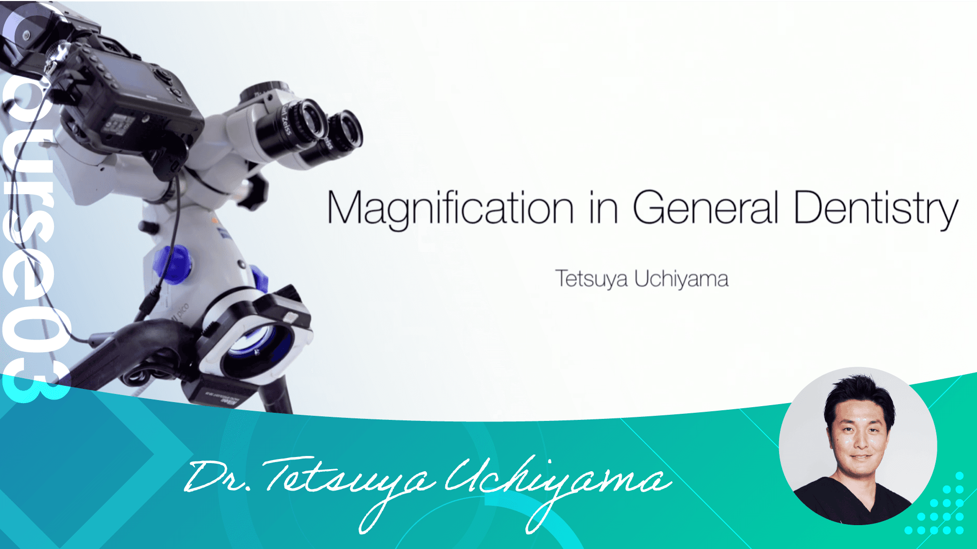
Dr. Uchiyama Course 03
Microscope
Microscope (All Videos)
Jan 19, 2023
Index
This video includes all of the following videos from Course03 Microscope.
[#1 Advantages of Microscopes]
Do you think that the microscope is a tool for specialists who use it for treatments performed by specialists? According to Dr. Uchiyama, it is a necessary tool for generalists who can use it for any kind of treatment.
He told us about his encounter with the microscope and the changes that have occurred in his practice and body since he started using it in his clinical practice.
He also talks about the method he actually uses in his clinical practice from each point of view on the issue of whether the mirror technique is better or the direct view is better.
[#2 Vision Control in EXTARO 300 ]
Although we tend to focus on the ease of use of microscopes, such as magnification and handling, it is also necessary to know about the type of light source. He explained in detail the differences in visibility between xenon, halogen, and LED light sources.
He said, "When you use a microscope, you can simply see and fill the root canal there and it will be cured. That's all there is to it. That's all there is to it," said Dr. Uchiyama. You should not miss the actual clinical video of a root canal, which is difficult to see in the dark, taken with a microscope.
He also talks about a difficult case of root canal filling of a mandibular molar.
[#3 How to record movies with EXTARO 300]
When recording photos or videos of a microscopic examination, not just any camera will do. It is necessary to understand the features of the microscope and the features of the camera equipment, and to choose a combination that makes the best use of both.
In this session, we talked about the functions and features of microscopes, as well as the minimum camera functions you should know.
[#4 Microscopes and rubber dam]
Don't you think that rubber dams are just for performance and don't affect your clinical practice with or without them?
There was a time when Dr. Uchiyama himself actually did not use them. However, he now uses rubber dams in all cases when he can. He summarized the advantages of using rubber dams and how to prepare them in advance.
He also discussed how to form the abutment teeth with a microscope and how to create a provisional restoration. Please also pay attention to Dr. Uchiyama's focus on how to eliminate bleeding from the gingiva.
[#5 Advantages of rubber dam 1]
When rubber dam protection is used in restorative treatment, it is necessary to drill holes with a certain degree of vigor. Dr. Uchiyama has made it more efficient by using the same drilling method for anterior teeth, molars, and root canal treatment. In the first half of the lecture, he talked about the drilling and fixing methods and the use of floss to fix the rubber dam firmly in place.
In the second half of the lecture, he discussed a difficult case in which the gingiva was black and transparent due to root staining, and esthetically restored the tooth using a microscope and a rubber dam.
[#6 Advantages of rubber dam 2]
In the first half of the presentation, he discussed tips for molar rubber damming to ensure a good target tooth while maintaining a good field of view. The method of fixation without the use of clamps is very useful and improves operability and field of view.
In the latter part of the lecture, he taught us how to directly construct the abutment without grinding or extracting the tooth for anterior teeth with low crown height diameters due to living teeth. The best results can be achieved by understanding the characteristics of the materials used and selecting and treating the appropriate cases.
[#7 Bonding with microscope]
This is a CR restoration using a microscope and rubber dam. Using actual microscopic cases, he taught us how to do plaque out and how to remove excess resin, and he explained each step in detail, mentioning recommended instruments and materials.
When extraction is performed under the microscope, it is easy to see the difference between the bone surface, soft tissue, and even the defective granulation during curettage of the extraction socket.
This video shows the importance of preoperative CT diagnosis and the importance of anticipating post-extraction treatment.
